As an Amazon affiliate, I may earn from qualifying purchases. Please read our Disclaimer and Privacy Policy.
Looking for interesting facts about x-rays? I’ve got you covered. Keep reading and discover everything from the origins of x-rays to how they were once used in shoe stores.
I’ve put together these facts about x-rays for anyone with a genuine interest in their amazing origin and contribution to medicine. All of my sources are noted at the end of the post.
What’s the big deal about x-rays?
If you’ve ever had an x-ray (and there’s a good chance you have) you’ll know how easy and painless it is.
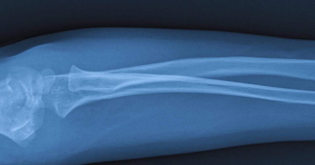
They can help locate broken bones, infections, or the presence of tumors in the body.
X-rays are one of the most powerful tools in modern medicine, allowing us to peer inside the human body and diagnose various conditions with remarkable precision.
But beyond their common use in healthcare, X-rays have a rich history and a variety of fascinating applications.
Of course, there are some limitations.
X-rays aren’t as effective at detecting tumors in soft tissues like the brain, liver, or muscle. In fact, small or early stage tumors may not be visible on x-ray at all.
As you can see there are plenty of interesting facts about x-rays and we’ve only just started. Keep reading for 15 interesting facts of x-rays.
1. Fun Facts On The Origin Of X-Rays
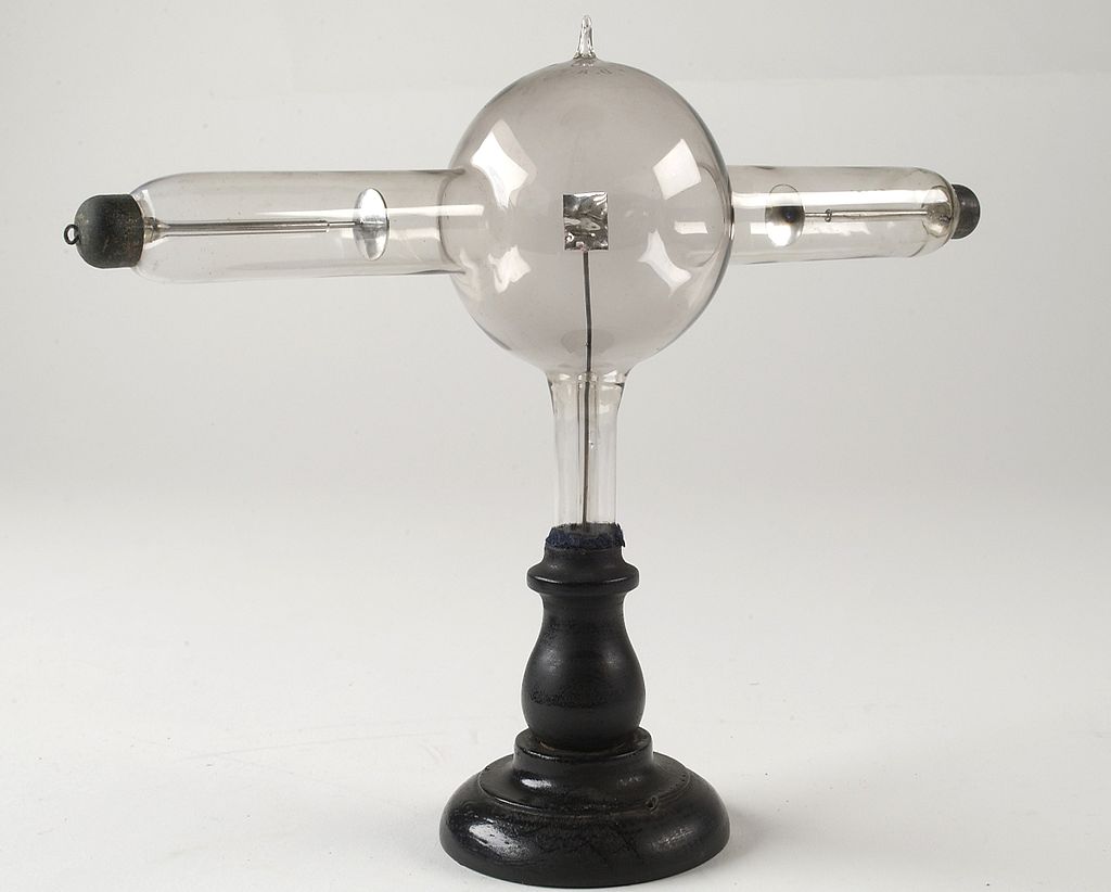
Did you know that x-rays were discovered by accident? It was 1895 when German physicist Wilhelm Corad
Röntgen was experimenting with cathode rays, streams of invisible electrons that are emitted through a vacuum tube.
As he experimented, he noticed something unusual.
A fluorescent screen in his lab began to glow even though it was shielded from the cathode rays.
This mysterious radiation, which he called “X” rays (because he didn’t know what else to call it), eventually led to the first Nobel Prize in Physics in 1901.
For reference, a vacuume tube is an electronic device that controls the flow of electric current in a high-vacuum environment.
They were widely used in the first half of the 20th century in radios, televisions, computers, and other electronic devices.
Although they were largely replaced in the technology noted above, they are still used in x-rays today. Modern vacuum tubes are designed with much greater precision.
2. Weird Ways X-Rays Were Used – Interesting Facts About X-Rays
I think it’s safe to say we all know the importance of well fitted shoes. But people in the early 20th century took it to a whole other level.
At the time, x-rays were so new and exciting that it seemed like a good idea to put them in shoe stores. The excuse was to make sure people’s shoes fit properly.
I suspect they, like me, just loved new gadgets. Unfortunately, these shoe-fitting fluroscopes proved to be a bad idea.
They certainly allowed the general public to have a look at what their foot bones looked like, although it was questionable as to how much it actually helped people fit their shoes.
By the 1920s, many X-ray pioneers suffered well-publicized, painful, and often gruesome deaths due to excessive radiation exposure.
Although a hard lesson to learn, it quickly become clear that conventional x-ray machines offered customers little more than excessive radiation exposure.

3. Helping Art Historians Uncover Secrets
Did you know that art historians can use x-rays analyze the layers of materials in a painting or sculpture?
They can also help identify the presence of foreign materials, such as repairs or additions made to the artifact.
X-rays can be used to determine authenticity, structural integrity, and material composition of works of art, like paintings.
Advances in x-ray technology have improved its application. Now, they use techniques such as x-ray fluorescence (XRF) and computed tomography (CT), which offers even more details
4. X-rays and the Discovery of DNA
X-ray crystallography is a specific application of X-ray technology that played a crucial role in the discovery of DNA.
Rosalind Franklin was a key figure in the discovery of DNA’s structure through her X-ray crystallography work.
Her X-ray images provided critical insights into the helical structure of DNA.
James Watson and Francis Crick, both DNA researchers, used Rosalind’s insights into x-ray diffraction data to build their model of DNS.
Together, they proposed that the DNA is structured as a double helix with two strands running in opposite directions, held together by base pairs.
This breakthrough explained how genetic information is stored and transmitted.
5. A Look Inside the Human Body – Interesting Facts About X-rays
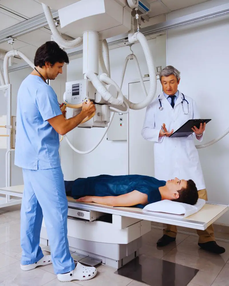
One of the most common uses of X-rays today is in healthcare, where they provide a picture of the inside of your body.
Whether it’s a chest X-ray to check for lung cancer or a scan to detect bone fractures, X-rays are invaluable for diagnosing a wide range of medical conditions.
If you’re into competitive sports, there’s a chance you’ve pulled a muscle or even fractured a bone at some point. Read: 35 Competitive Sports for Adults Over 50
6. How Many X-Rays Can You Safely Get?
According to the US Food and Drug Administration, there’s no specific number of x-rays a person can get before it becomes dangerous.
Every year 7 out of 10 Americans will need a medical x-ray or dental x-ray.
Sometimes, however, x-rays are taken when they’re not medically required.
Although today’s x-ray scans emit only a small amount of radiation, it may add slightly to the chance of getting cancer in later life.
In addition, changes can occur in the reproductive cells if the x-ray beam is on or near the sex organs.
The bottom line is only get x-rays when they are medically necessary. In some cases, the risk is worth the gain because they could save your life.
6. X-rays in Space: The Chandra X-ray Observatory
There are two types of x-rays associated with space. First, there are the naturally emitted x-rays from space. Specific celestial sources that naturally x-rays include:
- Black holes
- Neutron Stars and Pulsars
- Supernova Remnants
- Galaxy Clusters
- Quasars and Active Galactic Nuclei (AGN)
In order to study these naturally emitted x-rays, the United States of America launched the Chandra X-ray Observatory on July 23, 1999.
For more information on the Chandra Observatory, read: Observing the Universe in X-ray Light, published by the European Space Agency.
Chandra is one of NASA’s Great Observatories, a program that also includes the Hubble Space Telescope, the Compton Gamma Ray Observatory, and the Spitzer Space Telescope.

7. Digital X-rays: The Modern Evolution
Unlike the early days when X-rays were captured on photographic plates, modern digital X-rays use sensors to capture images that can be instantly viewed on a computer.
This technology reduces the amount of radiation needed and provides clearer images, enhancing the ability to diagnose conditions accurately.
More interesting facts about x-rays, particularly modern medical x-rays, include the following:
- Digital x-ray sysatems require less radiation to produce an image compared to traditional x-rays.
- They allow for adjustments in contrast, brightness, and zoom, helping to make more accurate diagnoses.
- Images are clearer and easier to read.
- The results are delivered faster.
- Digital x-rays can be integrated directly into a patient’s electronic health records.
- They can also be stored in the cloud.
- There is no need for chemical processing, unlike film-based x-rays.
- Digital x-ray technology has enabled the development of portable x-ray machines.
8. Radiation Dose: How Much is Too Much?
While X-rays involve exposure to electromagnetic radiation, the amount used in medical imaging is typically very low.
There is specifically identified number of x-rays that are considered bad for you. Ultimately, it’s best to only get x-rays when there is a medical benefit in doing so.
However, higher doses of radiation can have biological effects, which is why healthcare providers carefully control and monitor the use of X-rays, especially during procedures that require multiple scans.
9. From War to Peace: X-rays in Battlefield Medicine (Interesting Facts About X-rays)
During World War I, X-rays were used extensively to locate foreign objects like bullets in wounded soldiers.
This application of X-ray technology saved countless lives by allowing surgeons to safely remove shrapnel and other objects lodged in the body. It’s no surprise that gunshot wounds were often mentioned as a cause of injury.
10. Dental X-Rays
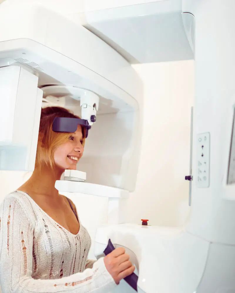
There are different types of x-rays that can be taken inside the mouth, called intraoral x-rays, or outside the mouth, called extraoral x-rays.
If you’ve ever had a dental x-ray, you may have been given a heavy lead apron to wear while the procedure is being done.
This is just a precaution. For women, use of the leaded apron will protect you and your fetus should you be pregnant.
If you are worried about radiation exposure, you may be reassured to learn that the amount of radiation in a single x-ray is very small.
11. The Many Types of X-rays
There are several forms of X-rays, each tailored to different medical, scientific, and industrial applications.
The main types include conventional X-rays, CT scans, digital X-rays, fluoroscopy, mammography, interventional radiology X-rays, X-ray fluorescence (XRF), and X-ray crystallography.
Each type uses X-ray technology in specific ways to provide valuable diagnostic and analytical information.
12. Benefits of Xrays
X-rays offer numerous benefits across various fields, particularly in medicine, dentistry, and security.
- They provide a non-invasive way of looking inside the body to help doctors diagnose certain issues.
- They can offer early detection of potentially serious conditions.
- Dental x-rays help detect cavities, bone loss, and other issues not visible during a regular examination.
- Specialized x-rays can help detect tumors and cancerous growths.
- X-rays also have a place in veterinary medicine where they are used to diagnose injuries, detect diseases, and monitor the health of animals.
- As mentioned earlier, x-rays can also be used to study artifacts.
13. John Hall-Edwards
John Hall-Edwards, a pioneering British surgeon, is recognized as the first medical practitioner to use X-rays for medical purposes.
It was just weeks after Wilhelm Röntgen’s discovery of X-rays when his colleague accidentally slipped a needle into his hand. It occured to him that he could use an x-ray to locate the needle quickly and it worked!
This event lead to the further use of x-rays in medical practice. Unfortunately, Hall-Edwards got a little to excited about x-rays and used them so often that he developed severe radiation burns.
The burns eventually lead to the amputation of his arm. Hard lesson learned.
14. Healthcare Worker protection
Health care workers protect themselves from radiation exposure by using PPE, such as lead aprons, lead gloves, and lead shields, to block harmful rays.
They rely on radiation monitoring devices to track exposure levels, maintain safe distances, and use strategic positioning to minimize risk.
Shielding and barriers are also employed to protect against stray radiation.
Comprehensive training and education ensure that workers understand how to effectively use these protective measures in their daily practices.
15. Marie Curie’s Impact on Mobile X-ray medicine
Marie Curie was clever and resourceful. When World War I broke out in 1914, she recognized the need to be able to make quick and accurate diagnoses on the battlefield.
Her idea was to put x-ray machines on wheels to make them mobile enough to get around quickly. The units, later known as “radiology cars” or “petites Curies” were mounted on cars and could be driven to the front lines.
Summary
rays have come a long way since their discovery over a century ago, transforming from a mysterious form of radiation into a vital tool across various fields.
Whether it’s diagnosing a broken bone or exploring the mysteries of the universe, the versatility and impact of X-rays are truly remarkable.
SOURCES:
National Institute of Biomedical Imaging and Bioengineering
Baring the Sole: The Rise and Fall of the Shoe-Fitting Fluoroscope

